Sanger Sequencing
Sanger Sequencing
by Hoi Kiu Wong

Creator: ktsimage | Credit: Getty Images/iStockphoto
Copyright: ktsimage
Introduction
DNA Sequencing has revolutionised Genomic Medicine, and has offered new and potential treatments to cancer patients, as well as towards other genetic diseases. With continuous development in the field of Bioinformatics, accuracy and precision in DNA Sequencing has improved drastically; from Sanger Sequencing to all kinds of Next-Generation Sequencing technologies with machines like Illumina and SOLiD System. In this article, we will outline what Sanger Sequencing is. In the next article (to be posted next week), we will look into Next-Generation Sequencing, and how it will play a pivotal role in Medicine.
PCR
Before we look into Sanger Sequencing in detail, we first need to understand PCR - the Polymerase Chain Reaction, as they are important in the amplification of the DNA strands in Sanger Sequencing.
The first step is to heat the reaction so that the DNA denatures, meaning that the two strands of DNA separate. Then, Taq polymerase (an enzyme) helps synthesise two new strands of DNA that are formed along the template strands (the two original strands of DNA). Now that there is four strands of DNA, each strand will now create two new copies - and the whole process repeats. This continuous cycle repeats approximately 30 to 40 times, which as a result produces more than one billion copies of the original DNA strand of interest (See Fig. 1). PCR is an automated cycling procedure and can be done in a few hours [1].

Fig. 1 Polymerase chain reaction (PCR)
Source: https://en.wikipedia.org/wiki/Polymerase_chain_reaction
Sanger Sequencing
Sanger Sequencing was first discovered by Frederick Sanger and his colleagues in 1977, and this technique is also known as the ‘chain-termination method’ of DNA sequencing. It works by replicating DNA during which dideoxynucleotides (ddNTP) would randomly incorporate itself into the DNA chain being synthesised, and this in turn would stop the chain from elongating [2]. This method uses Chain termination PCR (Polymerase Chain Reaction) in conjunction [3] - which is a technique that makes millions of copies of a particular section of DNA. PCR is important in Sanger Sequencing as many DNA copies are needed for the computer to be able to detect the oligonucleotides with high precision and accuracy [4].
To do this, a single primer that is complementary to the template DNA strand, starts a DNA synthesis reaction from its 3’ end. When this begins, DNA polymerase adds free deoxynucleotide bases to the growing DNA strand being created according to complementary base pairing. In order for this to occur, the reaction mixture has dNTPs (free deoxynucleotide bases), but they also have four different ddNTPs in the reaction mixture, one for each DNA base: A, T, C and G. These ddNTPS are artificially modified nucleotide bases - they are different to normal nitrogenous DNA bases as they lack a 3’ hydroxyl group which is required for further DNA chain elongation (more specifically, required for phosphodiester bond formation [3]), meaning that it will cause the termination of the DNA chain; and it is fluorescently labelled (has a fluorescent dye attached to it) which allows for detection of the DNA sequence later on in electrophoresis [5]. Note that Sanger Sequencing can be either manual or automated, the difference being that in manual Sanger sequencing, four PCR reactions are set up, each one with a single type of ddNTP (ddATP, ddTTP, ddGTP and ddCTP); whereas in automated Sanger sequencing, all ddNTPs are mixed into a single reaction, and each of the four dNTPs has a unique fluorescent label [3].
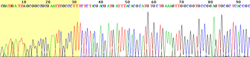
Fig. 2 An electropherogram created by Sanger Sequencing Method showing the base sequence of the DNA segment of interest
Source: Wikimedia Commons
https://commons.wikimedia.org/wiki/File:Sanger_sequencing_read_display.png
Detection of the DNA sequence uses electrophoresis [5]. The Sanger Sequencing method creates many copies of DNA fragments with different lengths and each one has fluorescent ddNTP at the end; these chains can also be called oligonucleotide copies of the DNA sequence [3]. These different DNA chains in the reaction mixture are then loaded to the sequencing machine (manually onto slab gels or automatically with capillaries) [5]. They are then electrophoresed which separates these DNA chains according to their length - to do this, an electric current is supplied, which causes these oligonucleotides (DNA chains of different lengths) to become negatively charged. As opposite charges attract, these oligonucleotides will be pulled towards the anode (positive electrode) on the opposite side of the gel (which is on the other side of the capillary tube). As all the oligonucleotides have the same charge per unit mass, their speed will be determined by the mass or size [3]. The shorter fragments (also known as the light bands or short chains) will travel further as they will experience less friction as they move through the gel matrix (in the capillary tube), thus they will pass through the laser beam first and are the first ones to be detected [3] [5].
The laser excites the fluorescent tags and causes a specific colour of light to be emitted (a specific wavelength emitted) - green for Adenine, red for Thymine, blue for Cytosine and yellow for Guanine, which are detected by the computer [6]. Then, the longer chains are later detected - as a result, the oligonucleotides will be arranged from smallest to largest, showing the order of the bases in the gene. In manual Sanger sequencing, note that there are four different PCR reactions thus there will be four separate lanes of gel; whereas in automated Sanger sequencing, there is only one. Once all the data has been obtained by the Machine, it can then be analysed and the computer generates an electropherogram (See Fig. 2) [5]. Since DNA Polymerase only synthesises DNA in the 5’ to 3’ direction starting at the primer, each ddNTP at the end will match with the nucleotide in the original sequence. Thus, by looking at the order of the bases from the shortest to the longest oligonucleotides, it is the same order as the DNA sequence going from the 5’ end to the 3’ end [3]. See Fig. 3 for a summary of automated Sanger Sequencing.

Fig. 3 Sanger Sequencing
Source: https://zhonglab.gitbook.io/3dgenome/chap0-preparation/0.2-sequencing-technologies
Bibliography
[1] “Polymerase Chain Reaction (PCR) Fact Sheet.” Genome.gov, www.genome.gov/about-genomics/fact-sheets/Polymerase-Chain-Reaction-Fact-Sheet.
[2] Morganti S. et al. (2020) Role of Next-Generation Sequencing Technologies in Personalized Medicine. In: Pravettoni G., Triberti S. (eds) P5 eHealth: An Agenda for the Health Technologies of the Future. Springer, Cham. https://doi.org/10.1007/978-3-030-27994-3_8
[3] “Sanger Sequencing Steps: DNA Sequencing.” Sigma, www.sigmaaldrich.com/technical-documents/articles/biology/sanger-sequencing.html.
[4] “What Is PCR (Polymerase Chain Reaction)?” Facts, The Public Engagement Team at the Wellcome Genome Campus, 25 Jan. 2016, www.yourgenome.org/facts/what-is-pcr-polymerase-chain-reaction.
[5] Applied Biological Materials, director. 1) Next Generation Sequencing (NGS) - An Introduction. YouTube, 22 June 2015, www.youtube.com/watch?v=jFCD8Q6qSTM.
[6]“Contexo.Info I Weave Together Information.” DNA Sequencing | Contexo.Info, www.contexo.info/dna_basics/dna_analysis/dna_sequencing/.


Comments
Post a Comment