Bioluminescence: The Mystery Behind The Light
Bioluminescence: The Mystery Behind the Light
by Hoi Kiu Wong

INTRODUCTION
Have you ever taken a stroll along the beach and had something intriguing catch your eye? A microscopic creature, glowing in the pitch-black sea? Unfortunately, I have only seen this ‘scene’ in documentaries; but that was already enough to grab my attention. As I have researched Bioluminescence, I have discovered how essential it is in the lives of millions of different species - from insects like fireflies to jellyfish, the list is endless. But the question remains: what is the science behind it?
THE SPECIALTY OF BIOLUMINESCENCE
Bioluminescence is defined as ‘the emission of light from living organisms (such as fireflies, dinoflagellates, and bacteria) as the result of internal, typically oxidative chemical reactions’[1]. Hence, this light produced by the result of these ‘chemical reactions’ is what sets Bioluminescence apart from other natural optical phenomena, such as fluorescence and phosphorescence. Fluorescent molecules, unlike bioluminescent molecules, do not produce their own light; they absorb photons, and in turn excite electrons to a higher energy state. As these electrons relax to their ground state, they re-emit their energy at a longer wavelength. This excitation and relaxation happens very quickly, therefore fluorescent light is only seen while the specimen is being illuminated [2] (See Fig.1). Similarly, Phosphorescence also re-emits light; however, it does so over a larger time scale, rather than immediately, and continues after excitation occurs (additional excitation) [3]. It is important to understand the distinction between fluorescence and bioluminescence because there is often confusion between them due to the fact that in some organisms, bioluminescent energy is used to excite fluorescence [4].

DISTRIBUTION OF BIOLUMINESCENCE
An apparent peculiarity of bioluminescence is that there is no obvious rule or reason in the distribution of luminous species among microbes, protists, plants and animals. Harvey (1940, 1952) expressed it in this way: “It is as if the various groups had been written on a blackboard and a handful of damp sand cast over the names. Where each grain of sand strikes, a luminous species appears. It is an extraordinary fact that one species in a genus may be luminous and another closely allied species contains no trace of luminosity.” Putting this into context, Cnidaria and Ctenophora have received the most sand; therefore, many members of the former phylum and nearly all of the latter are luminous, whereas there are certain phyla that contain no luminous organisms. There are also some cases in closely related genera of the same family where one genus is luminous while the others are not.
Another peculiarity of bioluminescence is that more bioluminescent organisms are marine creatures rather than terrestrial or freshwater inhabitants. There are very few non-marine organisms that are bioluminescent, hence the reason why they can easily be listed here: fireflies and beetles, earthworms, millipede Lumin-odesmus, limpet Latia, snail Quantula, the glow worms Arachnocampa and Orfelia, and luminous mushrooms. [12]
CHEMICAL STUDY OF BIOLUMINESCENCE
Bioluminescence has captured the interest of mankind ever since ancient times - descriptions of light emitted from fireflies can be found in folklore, songs and in numerous publications of literature; there is therefore no doubt that studies on Bioluminescence had already began in the early 17th century. However, its chemical study only originates from the early 20th century, when human research and technology began to become progressively more advanced [5].
LUCIFERIN-LUCIFERASE REACTION
Bioluminescence is produced by the oxidation of luciferin (known as the oxidizable substrate [8]) (Fig.2), and the rate of reaction can be controlled by a catalysing enzyme, either a luciferase or a photoprotein [2] such as aequorin [7] (a photoprotein found in 1962). This was first discovered when Dubois demonstrated the first example of a luciferin-luciferase reaction in 1885. He made two aqueous extracts from the luminous West Indies beetle Pyrophorus - one of which was prepared by crushing the light organs of the beetle in cold water, resulting in a luminous suspension. The luminescence gradually weakened and finally disappeared. The other was prepared in the same way but with hot water, which immediately put out the light, before being cooled. The two extracts produced light when mixed together. Dubois then repeated the experiment with the extracts of the clam Pholas dactylus and received similar results. Therefore, he concluded that the cold water extract contained a specific, heat labile enzyme necessary for the light-emitting reaction, and introduced the term ‘luciferase’ for this enzyme. He also concluded that the hot water extract contained a specific, relatively heat stable substance called ‘luciférine’ (presently spelled luciferin). Hence, the luciferin-luciferase reaction is an enzyme-substrate reaction that emits light [12].

Another person who made a significant contribution to the study of bioluminescence was E. Newton Harvey (1887-1959). In 1917, Harvey conducted experiments on bioluminescence but found that the light observed was weak and ‘short-lasting’. Following up in 1947, McElroy found that the light-emitting reaction requires ATP (adenosine triphosphate) as a cofactor(1), which was discovered in the firefly system [12]. Adding ATP to the mixtures of luciferin and luciferase resulted in a bright, long-lasting light. In fact, this was not a simple experiment at the time as ATP was not commercially available, thus this discovery was a huge breakthrough for the chemical study behind bioluminescence [5]. In 1949, McElroy and Strehler further found that luminescence reactions require another ion or cofactor Mg2+ (or Ca2+) in addition to luciferin, luciferase and ATP [5], which in turn causes a conformational change in the photoprotein, thus giving the organism a way to precisely control light emission [2].
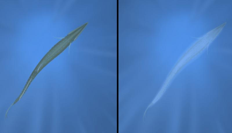
It was widely believed that bioluminescence was derived from luciferin-luciferase reactions, until the discovery of photoprotein aequorin in 1962. Aequorin emits light in aqueous solutions by the simple addition of Ca2+, regardless of the presence or absence of oxygen (Fig.3). The luminescence is emitted by an intramolecular reaction of the protein, the total light emission being proportional to the amount of the protein used. The definitions of luciferin or luciferase did not match up with the properties shown by aequorin, thus making it an exception at the time. However, in 1966, another bioluminescent protein in the parchment tube worm Chaetopterus was discovered, this one emitting light when a peroxide and Fe2+ are added; and similarly to aequorin, it was found that the total light
emission was proportional to the amount of the protein used. Hence, a new term ‘photoprotein’ was introduced for these unique bioluminescent proteins. (Shimomura and Johnson, 1966). A photoprotein could be an enzyme-substrate complex that is more stable than its dissociated components, enzyme and substrate. Due to its greater stability, a photoprotein occurs as the primary luminescent component in the light organs of luminous organisms instead of its dissociated components. In the light organs of Aequorea, aequorin is highly stable when Ca2+ is absent, but its less stable components, coelenterazine and apoaequorin, are hardly detectable in the jellyfish. [12]

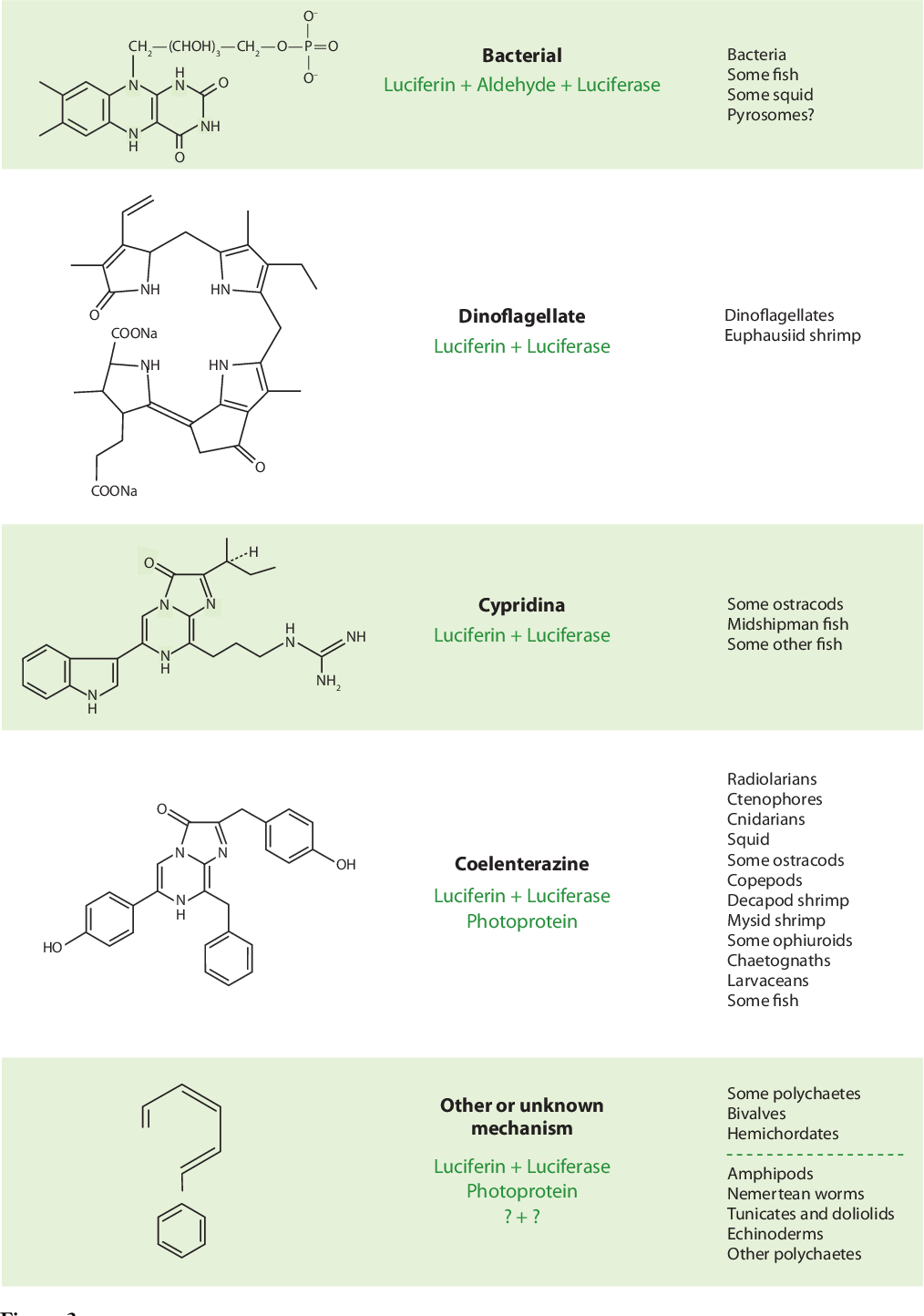
There is a large chemical variety of luciferins as many have derived from many evolutionary lineages [2], thus all of these bioluminescence reactions vary, except that they all require oxygen at some point. Harvey stated in his 1952 book “It is probable that the luciferin or luciferase from a species in one group may be quite different chemically from that in another” after he discovered that the luciferin of the clam Pholas differs from that of the ostracod Cypridina (Harvey, 1920). [12] However, this belief did not last long as it was discovered that a chemically identical luciferin can appear in unrelated organisms. For instance, around 1960, a luciferin identical to the luciferin of Cypridina was discovered in the luminous fishes Parapriacanthus and Apogon. Moreover, in the 1970s to the 1980s, it was discovered that coelenterazine luciferin is the light emitter in at least nine phyla: protozoans, jellyfish, crustaceans, molluscs, arrow worms and vertebrates. The likely explanation for this is that luciferin is acquired exogenously(3) through the diet. Hence, they are relatively easy to obtain as they are present in both luminous and non-luminous marine animals. However, the complete biosynthesis pathway is still not completely understood for any marine luciferins, so their ultimate origins are still unknown. [2] (Fig 4.)
Furthermore, delving deeper into the aspect of its chemical structure, some investigators found that highly purified extracts of luciferin contains a -COCH2OH side chain, which is oxidatively degraded to -COOH in the luminescent region. Hence, Cypridina luciferin is readily oxidized by many oxidizing agents, but produces light only when oxidized in the presence of luciferase [8].
It is still not known how many types of luciferin there are, but those that are better-studied are D-luciferin (found in fireflies), coelenterazine (most widely used luciferin in the sea) and Vargulin/Cypridina luciferin [2] (See Fig. 3).
BIOLUMINESCENT ORGANISMS
Bioluminescence spans in a range of ecosystems - the most comprehensive list of bioluminescent genera, assembled by Herring (1987) and Harvey, reports that of the seventeen phyla in the animal kingdom, at least eleven contain luminous forms [8]. This property of luminescence may be emitted by marine animals either by their luminescent organs or the bacteria on their bodies. Previously, there were many speculations that this light is produced by bacteria; however, modern scientists and researchers have proven that the bacteria emit light only after they have developed exponentially on dead fish and other organisms. Therefore, often, bioluminescence in the sea is usually due to large numbers of jellyfish, snails, fish, dinoflagellates and many more [8].
Dinoflagellates
Alongside fireflies, dinoflagellates are one of the most commonly encountered bioluminescent organisms. They are microscopic in size (range from about 30 μm to 1mm) and are often found in coastal regions [18], where a large number of them create specks of light seen in the water. Therefore, they create red tides, the phenomena in which the water is discoloured due to the high abundance of them (the dinoflagellates) rapidly growing and accumulating. At night,‘Bioluminescent bays’ are the result of the bioluminescent dinoflagellates creating a beautiful ‘sparkle’ along the beach and they have become famous tourist destinations in Puerto Rico and Jamaica [2].
FUNCTIONS OF BIOLUMINESCENCE
During the day, sunlight filters down into the ocean water, increasing the visibility. This means that many marine creatures would be much more vulnerable to predators as there are no hiding places at all in the shallower water, hence many would swim deeper into the ocean (approximately 1000m below the water surface) where visible light is unable to reach them. Therefore, this results in massive animal migration patterns in the planet’s oceans - animals vertically migrate upwards during the night when it is dark so they can look for food, and vertically migrate downwards when there is daylight in order to hide. [10]
As a consequence of this migration, most of these creatures spend a lot of time in dim light or in total darkness. Thus, bioluminescence helps them with their survival in different ways. [10]
To attract prey or to locate prey
Bioluminescence can be used by marine organisms to attract prey. An example of this is that some fish have red-emitting light organs located under their eyes, and the unusual long-wavelength sensitivity of the fish eye suggests that their red luminescence may be used to illuminate prey that are blind to red light, thus helping them hunt down prey. [2] The Angler fish is another example of a marine organism using bioluminescence to attract prey - many species of them have a small glowing bulb known as the esca (the “lure”) dangling in front of their mouth which contains tiny luminescent bacteria called photobacterium, hence they are able to attract prey and attack it. This creates a ‘win-win situation’ for both the anglerfish and the bacteria because the anglerfish is able to lure in prey and in exchange, the bacteria gains protection and nutrients from the fish as it is its host [15].
To act as a defense against predators
This is one of the most common functions of bioluminescence in the sea. Many marine creatures such as crustaceans, squid, jellyfish and fish release their light-emitting chemicals into the water, producing clouds or particles of light that distracts or blinds the predator (known as a ‘smokescreen’). Some even squirt their predators with luminescent slime, making them easy targets for secondary predators. Another way that these organisms use their bioluminescence when attacked is to lure in the secondary predators, thus giving them an opportunity to escape, as the first attacker would try to escape as well.
Some organisms also use their bioluminescence as a warning to predators, signalling the unpalatability(2) of the prey. [9]
Camouflage
Counterillumination is a process in which some marine organisms such as the hatchet fish or the poryfish use to camouflage themselves. The silhouette of the opaque animal is replaced with bioluminescence of a similar colour to the background of the ocean (blends in with the light filtering from the sky). This is most common amongst fishes, crustaceans, and squid that inhabit the twilight zone of the ocean where many predators have upward-looking eyes adapted to locate silhouettes of prey. [10]
Keeping the school together
Luminescent shrimp, squid and fish form schools, and many of them show vertical migration during the day and night. Thus, the luminous flashes from these marine organisms help keep the school together, as the light they emit can be detected over large distances. Dennell (1955) believed that the light of bathypelagic decapod crustaceans could be seen at distances up to 100m. Moreover, it is likely that luminescence is subject to diurnal rhythmicity and that the members of a school may mutually stimulate each other i.e. a luminescent euphausiid or a myctophid flashes light, and other individuals of the species may flash in turn as they are stimulated by the luminescence. Hence, it is widely believed that the light helps with regulating the degree of rising and sinking that the school needs to execute. Kampa & Boden (1957) found that flashing became most frequent during twilight migration, and the mean intensity, 1 × 10⁻⁴ μW/cm2, equalled that of the light-level with which the migration of the animals was associated. [9]
Courtship
Fireflies are well known for their bioluminescence during the warm summer months as they use their light to attract members of the opposite sex [13]. This is done through means of species-specific spatial or temporal patterns of light emission. [9] This can also be seen in marine life; for instance, the male Caribbean ostracod, a tiny crustacean, uses bioluminescent signals on its upper lips to attract females; Syllid fireworms are inhabitants of the seafloor, but when there is a full moon, they move to the open water where the females of some species, like Odontosyllis enopla, use bioluminescence to attract males while moving around in circles. [17]
Some other functions of Bioluminescence are included in Fig. 7

LUMINOUS BACTERIA IN THE SEA
Luminous bacteria form specific symbioses with some marine fish and squid, and this creates a ‘win-win situation’ for both the bacteria and the marine organism. This is because the bacteria provides the host with light that can be used to attract prey and to find a mate, while the host provides the bacteria with an ideal growth environment. For free-living bacteria where the adaptive value is less evident, the most generally accepted hypothesis is that the luminous bacteria growing on fecal pellets may serve as an attractant, causing the pellets to be consumed and in turn introducing the bacteria to an animal’s nutritious stomach and intestine. [10]
APPLICATIONS OF BIOLUMINESCENCE
BBioluminescence doesn’t only help marine animals or fireflies, they are also used as analytical tools in various fields of science and technology. For example, firefly bioluminescence is used as a method of measuring ATP (vital for living cells) [12] - this is done by adding a known amount of luciferin and luciferase to a blood or tissue sample, where the cofactor concentrations may be determined from the intensity of the light emitted [13]. Moreover, Ca2+- sensitive photoproteins e.g. aequorin from a jellyfish, are used in monitoring the intracellular Ca2+ that regulates many important biological processes; and certain analogues of Cypridina luciferin are utilized as probes for measuring superoxide anion, an important but rare substance in biological systems. Furthermore, the green fluorescent protein (GFP), which was discovered alongside aequorin, is used as a highly useful marker protein in biomedical research [12].
Similarly to methods of measuring ATP, scientists have also used these bioluminescent reactions in quantifying other specific molecules that are not involved in the bioluminescence reaction. They do this by attaching luciferase to antibodies, and the antibody-luciferase complex is then added to a sample where it binds to the molecule to be quantified. Following washing to remove unbound antibodies, the molecule of interest can be quantified indirectly by adding luciferin and measuring the light emitted. Methods used to quantify certain compounds in biological samples such as the ones described here are called assays. [13]
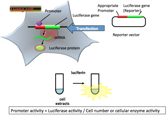
In fact, luciferases are good reporter enzymes that are used in the field of bioresearch. They are widely used in various aspects of biological functions, such as gene expression, post-translational modifications, and protein-protein interaction in cell based assays [16] (Fig. 6). This means that luciferases are used to study how individual genes are activated to code for protein or repressed to stop producing protein. The gene promoter is the genomic DNA sequence immediately upstream of the transcription start site [14], and this specific gene promoter can be attached to the DNA that codes for firefly luciferase and introduced into an organism. The activity of the gene promoter can then be studied by measuring the bioluminescence produced in the luciferase reaction. Thus, the luciferase gene can be used to “report” the activity of a promoter for another gene [13] (also known as quantitative visualization of gene expression).
In vitro and In vivo Bioluminescence Imaging
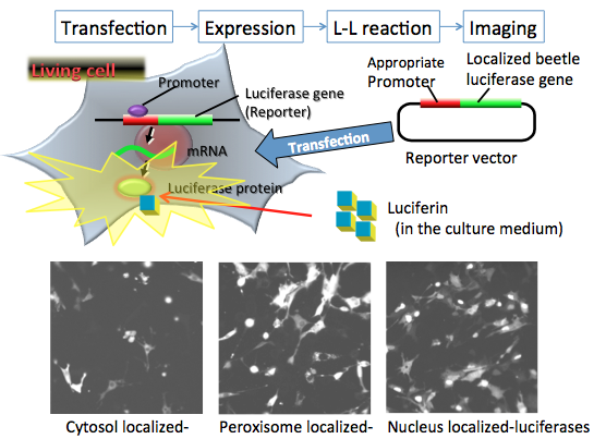
Molecular imaging using bioluminescent reporters is one of the most sensitive methods, and it has the cheapest and simplest procedure. Bioluminescence imaging using luciferase reporters does not need exogenous light illumination, and the luminescence reaction is quantitative. In In vitro bioluminescence imaging for organelles in living cells, after transfection of the reporter plasmid vector (consisting of the constitutive promoter sequence and the organelle targeting luciferase gene sequence for cytosol, peroxisome and nucleus in mammalian cells) into the target cells, the promoter region regulates the expression of luciferase genes in living cells. For the imaging experiment, firefly luciferin added to the medium enters into the organelle, where it is catalyzed by the expressed firefly luciferase to produce luminescence. The light signal can therefore show the locality or mobility of organelles in living cells. This is measured by special equipment using a CCD photon imaging system [16].
In vivo bioluminescence imaging is most commonly used for cell tracking. Luciferase-expressing cancer cells, immune cells, stem cells, or other types of cells can be imaged in small animals. After the reporter plasmid vector (consisting the constitutive promoter sequence, a luciferase gene sequence and antibiotic resistance sequence) is transfected into the target cancer cells, the promoter region regulates the expression of the luciferase gene in living cells. The luciferase expressed stable cells are then transplanted into the mouse. After allowing time for cancer cell growth, luciferin is injected into the body. Most commonly in these imaging experiments, firefly luciferin enters into the cells through the blood, where it is catalyzed by the expressed firefly luciferase to produce light. The light signals help show the location and size of cancer cells in the body (detected by CCD photon imaging system). Hence, providing information about the number and spatial distribution of the cells in the animal [16].

Furthermore, bioluminescence can be used to test for water purity. This can be done by placing genetically modified microorganisms into the water and their degree of luminescence can be used to identify certain toxins in the solvent. Many scientists have studied into this and have found that it is particularly effective in determining the presence of arsenic (a common water contaminant) and oil hydrocarbons [19]. Moreover, Bioluminescence can be applied in daily life; for instance, Portuguese fishermen have made use of the luminescent secretion of Malacocephalus for illuminating their bait, and their success gives hope that artificial luminous lures could work in the sea. [9]
In the future
Bioluminescent technology is still developing and scientists are trying to find innovative ways of using bioluminescence reactions. An idea for the future is that instead of using electricity, we could use bioluminescence to provide energy for our light sources. Bioluminescent algae would be stored in a long glass tube in salted water (creating a small ecosystem) and it would be able to lighten the surroundings. Also, many researchers are developing methods to create bioluminescent trees to line streets. This would effectively eliminate the need to place more expensive electrical lamps. Although the biggest challenge now is increasing bioluminescent brightness to provide enough light for drivers, I believe that these ‘glowing’ trees planted on the side of roads will become part of the norm within a few decades [19].
CONCLUSION
To conclude this article, I must admit that Bioluminescence is a very complicated phenomenon, especially since it requires studies spanning through morphology, cell biology, physiology, spectroscopy, organic chemistry, biochemistry and genetics. Therefore, the feeling of confusion is acceptable when reading about bioluminescence as many parts of this phenomenon are still unknown to mankind, thus further research into the topic is required. Although it is hard to understand completely, Bioluminescence is undoubtedly one of the most beautiful mysteries in nature that the world has ever seen. Not only has it helped numerous organisms with their survival, but it has already been helping us with advancing science and technology, even having the potential to save lives. Therefore, I believe that bioluminescence can assist mankind in innovation, and help shape our future significantly.

GLOSSARY
Cofactor: mostly metal ions or coenzymes, are inorganic and organic chemicals that assist enzymes during the catalysis of reactions.
Unpalatability: distasteful
Exogenously: originating from outside an organism.
BIBLIOGRAPHY
[1] “Bioluminescence.” Merriam-Webster, Merriam-Webster, www.merriam-webster.com/dictionary/bioluminescence.
[2]“Bioluminescence in the Sea.” Annual Reviews, www.annualreviews.org/doi/full/10.1146/annurev-marine-120308-081028. (website unavailable)
Haddock, Steven H.D., et al. “Bioluminescence in the Sea” . pdfs.semanticscholar.org/ae51/4348866380fa87daf2fdfa72b81c673fd391.pdf.
[3] “Fluorescence, Phosphorescence, Photoluminescence Differences.” Edinburgh Instruments, www.edinst.com/blog/photoluminescence-differences/.
[4]Monterey Bay Aquarium Research Institute (MBARI). “The Allure of Fluorescence in the Ocean.” YouTube, YouTube, 23 Aug. 2019, www.youtube.com/watch?v=whbeFXFZqiU&feature=youtu.be.
[5] “Bioluminescence: Chemical Principles And Methods.” Google Books, Google, books.google.com.hk/books?hl=en&lr=&id=yMLICgAAQBAJ&oi=fnd&pg=PR5&dq=bioluminescence&ots=ITJrdb8S_X&sig=-XdKtdlPhIm6YO3lLAGOEoK4wng&redir_esc=y#v=onepage&q&f=false.
[6] “The Bioluminescence Web Page.” The Bioluminescence Web Page, biolum.eemb.ucsb.edu/.
[7] Bhagat, Abhishek. “Harnessing Bioluminescence.” LinkedIn SlideShare, 2 Nov. 2016, www.slideshare.net/AbhishekBhagat17/harnessing-bioluminescence.
[8] Harleen Workman McAda. “Bioluminescence.” The American Biology Teacher, vol. 28, no. 7, 1966, pp. 530–532. JSTOR, www.jstor.org/stable/4441402.
[9] Nicol, J. A. “Bioluminescence.” Proceedings of the Royal Society of London. Series A, Mathematical and Physical Sciences, vol. 265, no. 1322, 1962, pp. 355–359. JSTOR, www.jstor.org/stable/2414178.
[10] Widder, E. A. “Bioluminescence in the Ocean: Origins of Biological, Chemical, and Ecological Diversity.” Science, vol. 328, no. 5979, 2010, pp. 704–708. JSTOR, www.jstor.org/stable/40655873.
[11] “Luciferase Reporters.” Thermo Fisher Scientific - US, www.thermofisher.com/hk/en/home/life-science/protein-biology/protein-biology-learning-center/protein-biology-resource-library/pierce-protein-methods/luciferase-reporters.html.
[12] Shimomura, Osamu. Bioluminescence: Chemical Principles and Methods. World Scientific Publishing Co. Pte. Ltd., 2019.
[13] MacKenzie, Steven. "Bioluminescence." The Gale Encyclopedia of Science, edited by K. Lee Lerner and Brenda Wilmoth Lerner, 5th ed., Gale, 2014. Gale In Context: Science, https://link.gale.com/apps/doc/CV2644030284/SCIC?u=hkharrow&sid=SCIC&xid=92707d85. Accessed 17 Oct. 2019.
[14] “Gene Promoter.” Gene Promoter - an Overview | ScienceDirect Topics, www.sciencedirect.com/topics/medicine-and-dentistry/gene-promoter.
[15] Ward, L.K. “Meet the Tiny Bacteria That Give Anglerfishes Their Spooky Glow.” Meet the Tiny Bacteria That Give Anglerfishes Their Spooky Glow, 8 May 2018, ocean.si.edu/ocean-life/fish/meet-tiny-bacteria-give-anglerfishes-their-spooky-glow.
[16] APPLICATIONS OF BIOLUMINESCENCE, photobiology.info/Ohmiya.html.
[17] Ocean Portal Team. “Bioluminescence.” Smithsonian Ocean, 18 Dec. 2018, ocean.si.edu/ocean-life/fish/bioluminescence.
[18] “Latz Laboratory.” Scripps Oceanography, scripps.ucsd.edu/labs/mlatz/bioluminescence/dinoflagellates-and-red-tides/dinoflagellate-bioluminescence/.
[19] Konica Minolta. “Living Light: Is There a Future For Bioluminescence Technology?” Konica Minolta Sensing Americas, sensing.konicaminolta.us/blog/living-light-is-there-a-future-for-bioluminescence-technology/.


Comments
Post a Comment