Medical Imaging
Medical Imaging
by Hoi Kiu Wong
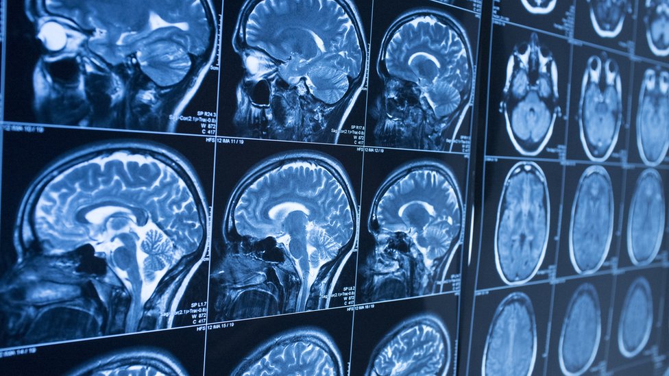
Image from Thinkstock
INTRODUCTION
In this modern era, technological advancements have integrated swiftly into our daily lives; giving us access to a vast amount of knowledge of the world, right under our fingertips. These developments have not only brought about self-driving cars and 5G, but also brought about improvements in the Medical field - especially in Medical Imaging. These include CT Scans, MRI Scans, PET Scans, and fMRI Scans; of which all are non-invasive. In this article, we will look into how they work and respectively, their useful functions.
COMPUTED TOMOGRAPHY (CT)
Computed Tomography (CT) Scans consist of a series of X-rays images taken from different angles around the body. These images are then processed by a computer which brings all the images together to produce cross-sectional images of the bones, blood vessels and tissues in the body [1]; therefore are more useful than regular X-rays scans that only produce a single 2D image. This medical imaging technique was first developed by South African American physicist Allan M. Cormack, and British electrical engineer Godfrey N. Hounsfield, and they were both awarded the 1979 Nobel Prize in Physiology or Medicine for this invention [2].
To carry out a CT Scan, the patient lies on a bed that moves through the gantry while the x-ray tube rotates around the patient (See Fig.1). It shoots narrow beams of x-rays through the body and these x-rays are then detected on the opposite side where the X-ray detectors are located. Dense structures (e.g. bones) absorb more radiation than less dense structure (e.g. soft tissues). Therefore, the denser structures show up lighter on the scan [3].

Fig. 1 Computed Tomography (CT) Scan
Creator: helovi
Image taken from: https://www.hopkinsmedicine.org/health/treatment-tests-and-therapies/computed-tomography-ct-or-cat-scan-of-the-brain
CT Scans have numerous advantages in regards to its ability to record X-ray images of the body at different angles, its quick speed, and its fewer restrictions on the mobility of the patient during the scan (in comparison to MRI Scans, where the patient needs to be completely still). However, there are also drawbacks to CT Scans - X-ray is a form of ionising radiation, so there is a higher risk of mutations in the body (in turn increases the risk of cancers); it can’t be used on Pregnant Women; the iodine-based contrasting agent (which is optional to take) may cause an allergic reaction, though this can be rare. Moreover, CT Scans are not as detailed as other medical Imaging Techniques such as MRI Scans, PET Scans and fMRI Scans [4].
MAGNETIC RESONANCE IMAGING (MRI)
Magnetic Resonance Imaging (MRI) Machines use powerful magnets and radio waves. Each machine has a magnet that is used to produce a strong magnetic field, which causes the protons in the body to align with the field. When a radiofrequency current is then pulsed through the patient, the protons are stimulated and spin out of equilibrium, straining against the pull of the magnetic field. When the radiofrequency current is removed, the MRI sensors can detect the energy released as the protons realign with the magnetic field, and the time it takes for them to do so. The data obtained from this allows physicians to differentiate between the various types of tissues according to these magnetic properties [5]. These MRI Scans are able to make very detailed cross-sectional images of the body (more detailed than CT Scan Images) (See Fig. 2). The 2D images can be put together to produce a 3D image after being processed by the computer. Moreover, MRI Scanner Machines look very similar to CT Scan Machines, as they both are tunnel-shaped.
MRI Imaging technique was developed by physicists Peter Mansfield and Paul Lauterbur, whom were jointly awarded the 2003 Nobel Prize in Physiology or Medicine for their discovery [6].
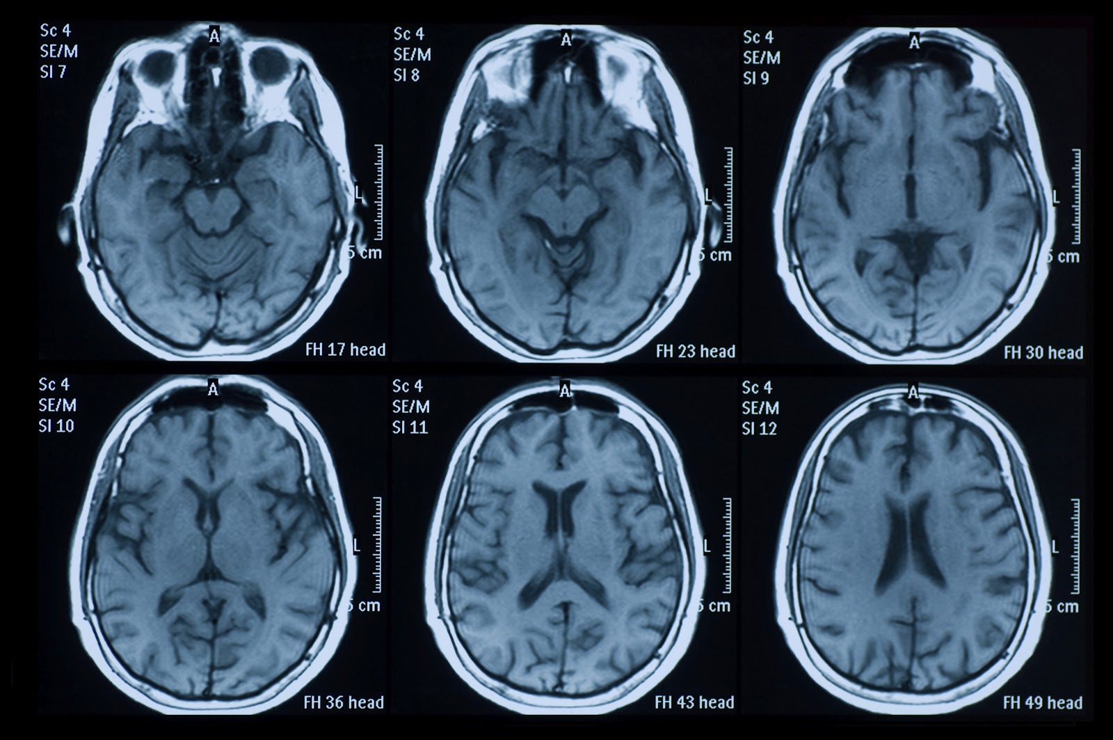
Fig. 2 MRI Scans of the Brain
Creator: temet
© temet
Credit: Getty Images
Image taken from: https://time.com/2860630/mri-scans-can-detect-early-onset-of-parkinsons-study-finds/
The main advantage of MRI Scans is that it doesn’t use ionising radiation; making it safe for pregnant women - over the past 30 years, there are no proven risks to pregnant women or unborn babies from MRI exams [7]; however, they aren’t usually recommended by doctors, particularly during the early stages of pregnancy [8]. Also, the images from MRI Scans are extremely clear and more detailed than images from other imaging techniques. The MRI Scans can also cover large portions of the body - so they are especially useful in determining whether a cancer has spread and assists doctors in determining the best treatment.
However, MRI Scan Machines are expensive. Though they may be able to detect cancers easily, they are unable to detect all cancers (i.e. breast cancers indicated by microcalcifications). It is also not possible to distinguish whether a tumour is malignant or benign (e.g. breast fibroadenomas) - which could lead to false positive results. Furthermore, patients who have claustrophobia should not get MRI Scans and there is a small chance of an allergic reaction to the contrasting agent received (or a skin infection at the site of injection). If a patient chooses to be sedated, there is a risk to that as well [9].
POSITRON EMISSION TOMOGRAPHY (PET)
Positron Emission Tomography (PET) Scans measure blood flow, oxygen, and sugar metabolism in the brain. It works by injecting a small amount of radioactive glucose (tracer) into a vein and the PET scanner then rotates around the body, detects the emission of radiation - providing information about where the higher levels of glucose are in the brain. Malignant tumour cells tend to show up brighter in images as they are more active than benign cancerous cells and absorb in more glucose than healthy cells [10]. Areas of high brain activity and metabolism may also be signs of Alzheimer’s (in particular, can highlight amyloid protein plaques in the brain [12]) or presence of seizures (See Fig. 3). As PET Scans look into activity of different parts of the brain, it can be used to investigate brain structure and function.

Fig. 3 PET Scans of a normal brain and a brain of a patient with Alzheimer’s disease.
Image taken from: [11]
https://www.mayoclinic.org/tests-procedures/pet-scan/about/pac-20385078
PET Scans are particularly useful in finding cancers and brain disorders. It is very often that physicians will combine PET Images with CT or MRI scans to have a detailed view of the body which can help lead to an accurate diagnosis Also, PET Scans can sometimes detect disease before it shows up on other imaging tests [11]. However, there are drawbacks to PET Scans - the patient has to be completely still, which limits the activities that can be investigated.
FUNCTIONAL MAGNETIC RESONANCE IMAGING (fMRI)
Functional Magnetic Resonance Imaging (fMRI) shows changes in the brain activity in real time. It does this by monitoring the supply of oxygen in the different regions of the brain. Regions of the brain that are more active will receive more oxygenated blood; hence, this technique heavily relies on the fact that cerebral blood flow and neuronal activation are linked [13]. The molecules in oxygenated blood respond differently to the magnetic field than those in deoxygenated blood; hence, the more active areas can be detected as they absorb less signals and emit less energy than less active areas [14]. Thus, these parts of the brain ‘light up’ (See Fig. 4).
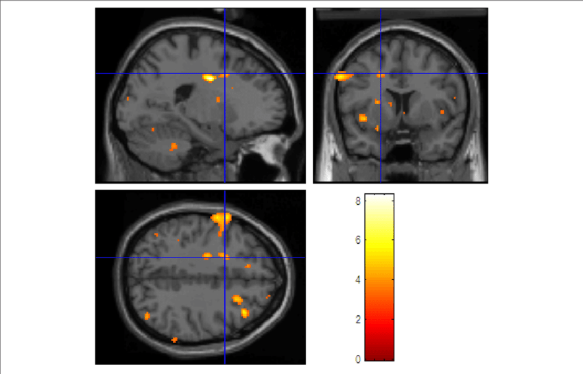
Fig. 4 fMRI Scans showing brain activity - yellow areas signify increased activity compared with a control condition.
Image taken from: Tian, Tian Siva. “Functional data analysis in brain imaging studies.” Frontiers in psychology vol. 1 35. 8 Oct. 2010, doi:10.3389/fpsyg.2010.00035
https://pubmed.ncbi.nlm.nih.gov/21833205/
Therefore, fMRI is particularly useful in investigating brain structure and function. However, one drawback is that the scanning procedure is very noisy and the patient needs to be completely still.
BIBLIOGRAPHY
[1] “CT Scan.” Mayo Clinic, Mayo Foundation for Medical Education and Research, 28 Feb. 2020, www.mayoclinic.org/tests-procedures/ct-scan/about/pac-20393675.
[2] “The Nobel Prize in Physiology or Medicine 1979.” NobelPrize.org, www.nobelprize.org/prizes/medicine/1979/summary/.
[3] “Computed Tomography (CT).” National Institute of Biomedical Imaging and Bioengineering, U.S. Department of Health and Human Services, www.nibib.nih.gov/science-education/science-topics/computed-tomography-ct.
[4] “Pros and Cons of a CT Scan.” Fairy Fire World, 20 Aug. 2017, www.fairyfireworld.com/pros-cons-ct-scan/.
[5] “Magnetic Resonance Imaging (MRI).” National Institute of Biomedical Imaging and Bioengineering, U.S. Department of Health and Human Services, www.nibib.nih.gov/science-education/science-topics/magnetic-resonance-imaging-mri.
[6] “The Nobel Prize in Physiology or Medicine 2003.” NobelPrize.org, www.nobelprize.org/prizes/medicine/2003/summary/.
[7] Radiological Society of North America (RSNA) and American College of Radiology (ACR). Patient Safety - MRI During Pregnancy, www.radiologyinfo.org/en/info.cfm?pg=safety-mri-pregnancy.
[8] “Who Can Have One - MRI Scan.” NHS Choices, NHS, www.nhs.uk/conditions/mri-scan/who-can-have-it/.
[9] “MRI.” CancerQuest, www.cancerquest.org/patients/detection-and-diagnosis/magnetic-resonance-imaging-mri.
[10] “What to Expect.” Stanford Health Care (SHC) - Stanford Medical Center, 12 Sept. 2017, stanfordhealthcare.org/medical-tests/p/pet-scan/what-to-expect.html.
[11] “Positron Emission Tomography Scan.” Mayo Clinic, Mayo Foundation for Medical Education and Research, 25 Aug. 2020, www.mayoclinic.org/tests-procedures/pet-scan/about/pac-20385078.
[12] Mann, Denise. “PET Scans May Help With Alzheimer's Diagnosis.” WebMD, WebMD, 11 July 2011, www.webmd.com/alzheimers/news/20110711/pet-scans-nay-help-with-alzheimers-diagnosis.
[13] “Functional Magnetic Resonance Imaging.” Wikipedia, Wikimedia Foundation, 7 Nov. 2020, en.wikipedia.org/wiki/Functional_magnetic_resonance_imaging.
[14] Watson, Stephanie. “How FMRI Works.” HowStuffWorks Science, HowStuffWorks, 7 Oct. 2020, science.howstuffworks.com/fmri.htm.


Comments
Post a Comment